 메뉴열기
메뉴열기
연구의 배경: X-선 발생장치를 이용한 관상동맥조영술 및 중재술은 허혈성심장질환을 진단하고 치료하는 주요치료지침이 된다. 일반적으로 X-ray발생장치를 이용한 영상획득은 방사선량이 많을수록 얻어지는 영상이 선명해지는 특징이 있기 때문에, 크기가 작은 심장혈관의 양질의 영상을 얻기 위해서 노출되는 방사선량은 필연적으로 다른 방사선학적 시술보다 양이 훨씬 많고 환자와 의료진 모두는 잠재적 위험요소에 노출 될 수 밖에 없다¹. 기존의 연구에서 심장중재시술의사들은 일반 심장내과의사보다 DNA의 손상 정도가 더 높다고 보고하고 있다². 방사선에 대한 인체의 생물학적 효과는 크게 피폭선량에 비례하여 그 영향이 나타나는 결정적 효과 (deterministic effect)과 그렇지 않은 확률적 효과 (stochastic effect)로 구별된다. 결정적 효과의 대표적인 문제는 피부, 골수, 생식세포의 억제등이 있고 다소의 논란이 있으나 백내장발생도 결정적 효과로 보는 것이 보통이다. 또한 확률적 효과로는 방사선 유발 암이나 유전학적 이상이 해당된다³. 정부는 방사선에 대한 환자의 임상적 안전기준을 갖추고 있으나 의료진에 대해서는 대부분 thermoluminescent dosimeters에 의한 흡수선량만을 기준으로 하고 있어 미흡하고, 특히 방사선량을 기준으로 볼 때 상대적으로 훨씬 많은 양을 차지하는 cineangiography에 대한 기준이 없이 fluoroscopy의 선량만을 기준으로 되어있고, X-선 발생장치에서 발산되는 선량에 대해서는 아무런 제한 기준이 없는 것도 문제라 할 수 있다4.
연구의 목적: X-선 발생장치에서 발산하는 선량을 Phantom cinegraphy를 이용하여 통상적 방사선량 프로토콜 (conventional radiation dose protocol, CDP)에 비해 절반 정도의 선량에 해당하는 저방사선량 프로토콜 (low radiation dose protocol, LDP)을 정하였다 (Table 1). 표준 관상동맥조영술을 시행받는 환자를 1:1 무작위 이중맹검의 방식으로 배정하여 시각적 진단에 영향을 미치지 않는 범위에서 관상동맥조영술을 시행후 방사선량 변수로 dose-area product (DAP) 와 air-kerma (AK) 값을 비교하였다. 유사연구가 없어서 본원에서 기존에 통상진료과정에서 사용된 저선량의 방사선량으로부터 표본수를 산정하여 양군에 50여명의 환자를 등록하기로 하였다.
Table 1. Radiation dose and frame rates setting according to the low and conventional dose protocol (LDP vs. CDP) in X-ray generating equipment of Artis-zeeTM (Siemens Health Care, München, Germany) monoplane coronary angiography system.
| LDP | CDP | |||
|---|---|---|---|---|
| Radiation dose | Frame/sec | Radiation dose | Frame/sec | |
| Fluoroscopy | 45 ηGy/frame | 10 | 45 ηGy/frame | 15 |
| Cineangiography | 0.12 μGy/frame | 10 | 0.24 μGy/frame | 15 |
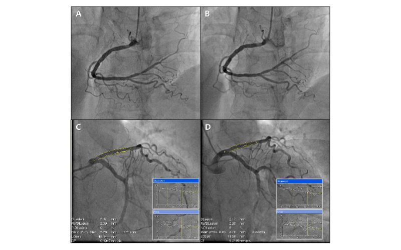
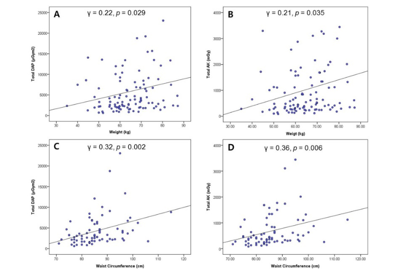
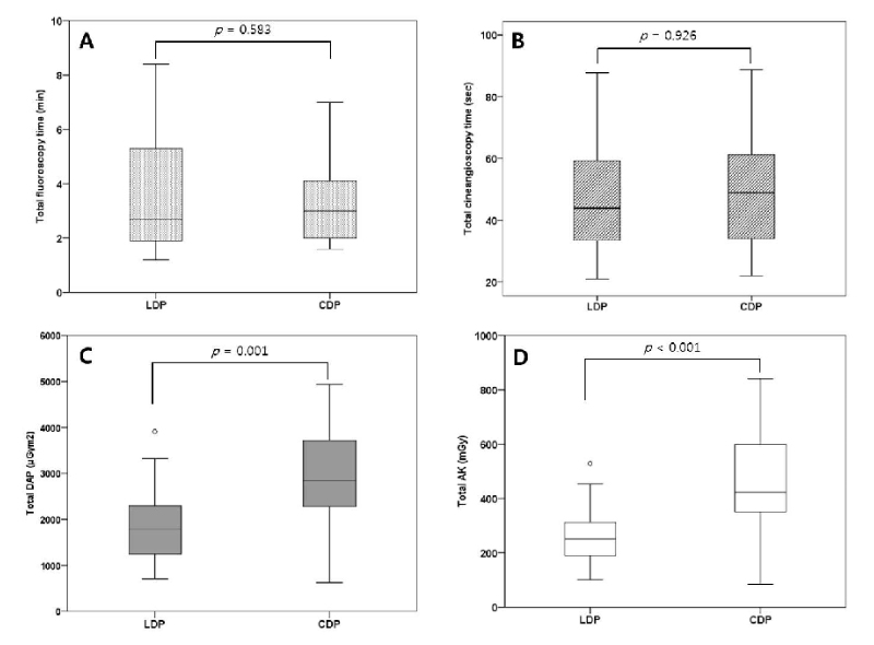
결과: 총 100여명 이상이 등록되었으며 관상동맥조영술 후 독립된 영상의사의 시각적 진단이 참조하였으며 양군에서 의미 있는 시각적 진단을 위한 방해요소는 발견되지 않았다 (Fig. 1). 장비에서 발산된 방사선량은 환자의 체중과 허리둘레에 비례하였다 (Fig. 2). 전체 방사선 노출시간, frame rate, 전체 정지영상의 수는 양군이 유사한 반면, LDP의 DAP와 AK 값은 CDP 선량보다 40%이상 적게 측정되었다 (Fig. 3).
Figure 1 A-D. Representative still image of coronary angiography taken by low and conventional radiation dose protocol (LDP vs. CDP). A. Right coronary angiogram with LDP in sixty eight years old women. B. Right coronary angiogram with CDP in same patient. C. Quantitative coronary analysis (QCA) of left coronary angiogram with LDP in eighty five years old women. D. QCA of left coronary angiogram with CDP in same patient.

Figure 2 A-D. The relationship between patient’s body size and estimated radiation dose. Body weight and waist circumference are well correlated while the body mass index and height are not (Data are not shown). A. Total dose area product (DAP) and weight. B. Air-kerma (AK) and weight. C. Total DAP and waist circumference. D. Total AK and waist circumference

Figure 3 A-D. Total fluoroscopic and cineangiographic time between LDP and CDP. A. Total fluoroscopic time (minutes) in LDP group are broader and mild higher without statistical significance than that of CDP. B. Total cineangiographic time (seconds) is not different. C and D. Estimated radiation dose (DAP and AK) according to the dose protocol.

고찰 및 결론: 기존의 연구를 토대로 볼 때 40%의 방사선량 감축은 시술과 관련된 환자와 의료진에게 방사선과 관련된 악성종양의 위험을 약 40% 감소시킬 수 있을 것으로 추정한다 [5]. 본 연구는 과거 연구들에서 방사선량 감축을 위해 여러 가지 원칙을 제시하였으나 장비에서 발생하는 선량을 줄여서 관상동맥의 검사나 시술에 적용한 무작위연구가 없다는 점에서 차별화 된다. 본 연구의 결과를 근거로 필요 이상의 과도한 선량을 발생하는 장비의 setting 값을 적절한 수준으로 조절 (optimization)하는 것은 환자뿐만 아니라 만성적으로 방사선 위해에 노출되는 의료진의 안전을 도모할 수 있는 전략을 제공할 수 있을 것으로 사료된다.
* Submitted to Juournal
References
1. Hirshfeld JW Jr, Balter S, Brinker JA, Kern MJ, Klein LW, Lindsay BD, Tommaso CL, Tracy CM, Wagner LK, Creager MA, Elnicki M, Lorell BH, Rodgers GP, Weitz HH; American College of Cardiology Foundation; American Heart Association/; HRS; SCAI; American College of Physicians Task Force on Clinical Competence and Training. ACCF/AHA/HRS/SCAI clinical competence statement on physician knowledge to optimize patient safety and image quality in fluoroscopically guided invasive cardiovascular procedures: a report of the American College of Cardiology Foundation/American Heart Association/American College of Physicians Task Force on Clinical Competence and Training. Circulation. 2005 ;111(4):511-32.
2. Sciahbasi A, Rigattieri S, Sarandrea A, Cera M, Di Russo C, Fedele S, Romano S, Pugliese FR, Penco M. Operator radiation exposure during right or left transradial coronary angiography: A phantom study. Cardiovasc Revasc Med. 2015 ;16(7):386-90.
3. Vañó E, González L, Beneytez F, Moreno F. Lens injuries induced by occupational exposure in non-optimized interventional radiology laboratories. Br J Radiol. 1998 ;71(847):728-33.
4. FDA Safety Advisory.
https://www.fda.gov/Radiation-EmittingProducts/RadiationEmittingProductsandProcedures/MedicalImaging/ucm115354. Accessed January 14, 2018.
5. Vijayalakshmi K, Kelly D, Chapple CL, et al. Cardiac catheterisation: radiation doses and lifetime risk of malignancy. Heart 2007 ;93:370-1.